Quick Answer
Dental X-rays are essential diagnostic tools that reveal problems invisible to the naked eye—decay between teeth, bone loss, abscesses, and tumors. Modern digital radiography reduces radiation exposure by 80–90% compared to film systems used decades ago. A full set of digital bitewings delivers roughly the same radiation as one day of natural background exposure. For most adults, bitewing X-rays every 12 to 24 months suffice if you have no active dental disease. High-risk patients—those with frequent cavities, gum disease, or dry mouth—may need them every 6 to 12 months. The risk of undetected disease far exceeds the minimal radiation risk from properly administered dental imaging.
What This Article Covers
-
Types of dental X-rays and what each reveals
-
Actual radiation exposure compared to daily life
-
Safety measures that protect patients
-
How often different patients need imaging
-
When to question or decline X-rays
-
What dentists look for on your films
Types of Dental X-Rays and What They Reveal
Dental imaging is not one-size-fits-all. Each type serves specific diagnostic purposes, and dentists select based on your symptoms, history, and examination findings.
Bitewing Radiographs
These show the crowns of upper and lower teeth in a single image, with special focus on the areas where teeth contact each other. Bitewings are the workhorse of routine dental surveillance.
What they reveal:
-
Decay between teeth that visual examination cannot detect
-
Bone levels supporting teeth, indicating early gum disease
-
Leaking or deteriorating fillings and crowns
-
Calcular deposits below the gumline
Frequency: Typically every 12 to 24 months for adults with stable oral health. Every 6 to 12 months for high-cavity-risk patients.
Periapical Radiographs
These capture the entire tooth from crown to root tip, including surrounding bone. Dentists use them to investigate specific problems rather than screen broadly.
What they reveal:
-
Abscesses or infections at root tips
-
Bone loss patterns around individual teeth
-
Root fractures or resorption
-
Cysts or other pathology in the jawbone
-
Status of teeth under development in children
Frequency: As needed based on symptoms or specific diagnostic questions. Not routine screening tools.
Panoramic Radiographs
A single image showing the entire mouth—teeth, upper and lower jaws, temporomandibular joints, and surrounding structures. The machine rotates around your head while you stand or sit still.
What they reveal:
-
Impacted teeth, particularly wisdom teeth
-
Jaw fractures and bone pathology
-
Sinus conditions related to upper teeth
-
Developmental abnormalities
-
Tumors or cysts in the jawbones
-
Temporomandibular joint overview
Frequency: Every 3 to 5 years for most patients, or before major dental procedures like implant placement or orthodontic treatment.
Cone Beam Computed Tomography (CBCT)
Three-dimensional imaging that provides detailed views of bone, teeth, and surrounding anatomy. CBCT delivers significantly more information than conventional two-dimensional X-rays.
What it reveals:
-
Precise three-dimensional position of impacted teeth
-
Bone volume and density for implant planning
-
Root canal anatomy before endodontic treatment
-
Fracture patterns and bone defects
-
Airway and sinus assessments
-
Tumor extent and relationship to vital structures
Frequency: Only when specific three-dimensional information is necessary. Not routine screening due to higher radiation than conventional radiographs.
Dr. Chan: “I explain CBCT to patients as the difference between a photograph and a CAT scan. For routine checkups, two-dimensional images are sufficient. When I’m placing an implant millimeters from a nerve, or evaluating a complex root canal, CBCT prevents complications that two-dimensional films miss. I use it when the diagnostic gain justifies the additional exposure.”
Radiation Exposure: Putting Dental X-Rays in Context
Patient anxiety about radiation often exceeds actual risk. Understanding comparative exposure helps make informed decisions.
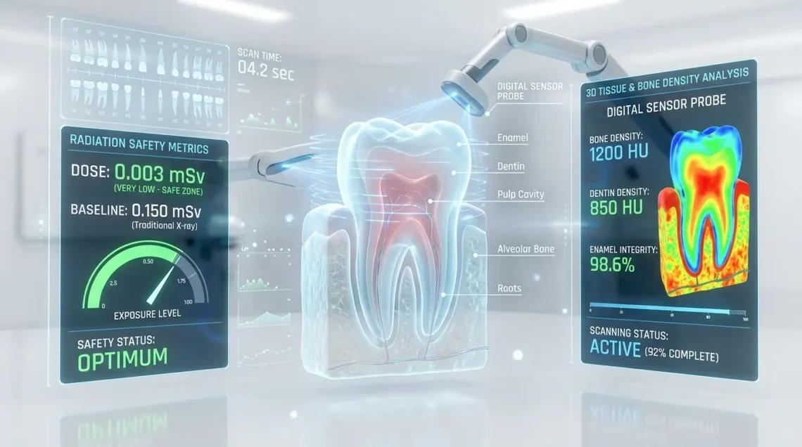
A single digital bitewing radiograph delivers approximately 0.005 millisieverts (mSv) of radiation. Four bitewings—the standard set—total roughly 0.02 mSv. For perspective:
-
One day of natural background radiation in the United States: approximately 0.01 mSv
-
A cross-country commercial flight: approximately 0.04 mSv
-
Chest X-ray: approximately 0.1 mSv
-
Mammogram: approximately 0.4 mSv
-
Abdominal CT scan: approximately 10 mSv
A full mouth series of 18 digital radiographs delivers approximately 0.09 mSv—roughly equivalent to nine days of natural background exposure or two cross-country flights.
Modern digital sensors require significantly less radiation than traditional film. Collimation limits the beam to the area of interest. Lead aprons with thyroid collars provide additional protection, though their necessity with digital systems is debated among experts.
The risk of missing a developing abscess, hidden decay, or early tumor far exceeds the theoretical cancer risk from dental radiography at these exposure levels. The American Dental Association and FDA both emphasize that radiation should be minimized, but not avoided when clinically necessary.
Safety Measures That Protect Patients
Dental practices employ multiple strategies to reduce radiation exposure while maintaining diagnostic quality.
Digital sensors replace film, requiring 70–90% less radiation for equivalent image quality. Immediate viewing eliminates retakes due to processing errors.
Rectangular collimation restricts the X-ray beam to the sensor size, reducing scatter radiation to surrounding tissues compared to round beams.
Lead aprons and thyroid collars shield radiosensitive organs. Thyroid collars are particularly important for children and pregnant patients.
ALARA principle—As Low As Reasonably Achievable—guides all radiographic decisions. Dentists take only the images necessary for diagnosis and treatment planning, selecting the fewest films that answer the clinical question.
Proper technique reduces retakes. Positioning devices and trained operators ensure first-attempt success.
Regular equipment inspection and calibration maintain optimal performance. State regulations mandate periodic testing of X-ray machines.
How Often Different Patients Need Imaging
Radiation frequency should match individual risk, not arbitrary schedules.
Low-Risk Adults
-
No cavities in past 24 months
-
No gum disease
-
No complex dental work
-
Good oral hygiene habits
Recommended interval: Bitewings every 24 to 36 months. Panoramic every 5 years unless symptoms arise.
Moderate-Risk Adults
-
One or two cavities in past 24 months
-
History of gum disease now stable
-
Multiple restorations requiring monitoring
-
Moderate oral hygiene
Recommended interval: Bitewings every 12 to 18 months. Periapicals as needed for specific teeth.
High-Risk Adults
-
Three or more cavities in past 24 months
-
Active or recurrent gum disease
-
Dry mouth from medications or medical conditions
-
High sugar or acid diet
-
Chemotherapy or radiation therapy to head and neck
-
Eating disorders with vomiting
Recommended interval: Bitewings every 6 to 12 months. Additional imaging as clinical situation demands.
Children and Adolescents
Developing dentition requires different monitoring than adult mouths. Eruption patterns, spacing for permanent teeth, and cavity risk in primary teeth guide frequency.
Recommended interval: Highly individualized based on cavity risk, age, and developmental stage. Typically every 6 to 12 months for high-risk children, extending to 12 to 24 months for low-risk adolescents with stable dentition.
Pregnant Patients
Routine dental X-rays are generally postponed until after delivery unless necessary for urgent diagnosis or treatment. When essential, abdominal shielding and collimation minimize fetal exposure. The American College of Obstetricians and Gynecologists confirms that dental radiography with proper shielding is safe during pregnancy when clinically indicated.
When to Question or Decline X-Rays
Informed patients participate in clinical decisions. You should understand why each image is recommended.
Appropriate questions:
-
What specific problem are you looking for with this X-ray?
-
How will this image change my treatment plan?
-
When was my last radiograph of this type?
-
Can you use my recent X-rays from another dentist?
Red flags suggesting overuse:
-
Full mouth series recommended at every cleaning without clinical justification
-
Refusal to explain why a specific image is necessary
-
No discussion of your individual risk factors
-
Outdated film-based systems in an era when digital is standard
That said, declining all radiography carries significant risk. Hidden decay between teeth, early bone loss from gum disease, and asymptomatic abscesses progress silently until they cause pain, tooth loss, or systemic infection. The cost of undetected disease exceeds the minimal radiation exposure from appropriate imaging.
What Dentists Look For on Your Films
Understanding the diagnostic value helps appreciate why X-rays matter.
Decay detection: Visual examination misses approximately 60% of interproximal cavities in early stages. Bitewings catch these when they are small fillings rather than root canals.
Bone level assessment: Gum disease destroys bone before teeth loosen. X-rays reveal this destruction in millimeters, allowing intervention before tooth loss becomes inevitable.
Root and nerve evaluation: Periapicals show whether decay has reached the pulp, whether cracks extend below the gumline, and whether infection has spread to surrounding bone.
Pathology screening: Panoramic films reveal cysts, tumors, and developmental anomalies that produce no symptoms until advanced. Early detection of oral cancers and odontogenic tumors dramatically improves outcomes.
Treatment planning: Implants, root canals, extractions, and orthodontics all require precise anatomical knowledge that only radiography provides. CBCT has become standard for implant planning to avoid nerve damage and ensure adequate bone volume.
The Bottom Line
Dental X-rays are safe, essential, and far lower in radiation than most patients assume. Modern digital technology has transformed the risk-benefit equation decisively in favor of appropriate imaging. The danger lies not in taking X-rays when indicated, but in refusing them and allowing silent disease to progress unchecked.
Discuss your individual risk factors with your dentist. Ask why specific images are recommended. Share previous radiographs to avoid duplication. But trust that when your dentist recommends imaging, it is because the diagnostic information protects your health in ways that visual examination alone cannot.
Your teeth and jawbone hide secrets. X-rays reveal them before they become emergencies.
Sources
-
American Dental Association. “Dental Radiographic Examinations: Recommendations for Patient Selection and Limiting Radiation Exposure.” ADA/FDA Guidelines, 2025.
-
Chan A. “Digital Radiography in General Practice: Patient Dose Reduction and Diagnostic Efficacy.” General Dentistry, 2025;73(4):389-395.
-
American Academy of Oral and Maxillofacial Radiology. “Selection Criteria for Dental Radiography: 2024 Update.” Oral Surgery, Oral Medicine, Oral Pathology and Oral Radiology, 2024;138(3):245-258.
-
Ludlow JB, et al. “Effective Dose of Dental CBCT: A Meta-Analysis.” Dentomaxillofacial Radiology, 2024;53(4):202-210.
-
White SC, Pharoah MJ. “Oral Radiology: Principles and Interpretation.” 9th Edition, Elsevier, 2025.